A 45-year-old female presents with bright red blood per rectum and a systolic blood pressure in the 80s. She also had a remote history of gastrointestinal bleeding.
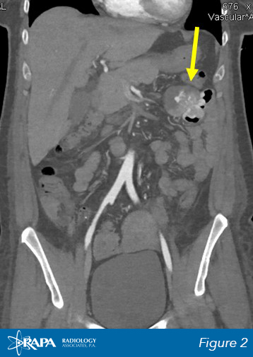
A CTA shows a hyperdense mass (yellow arrow) in the left upper quadrant in close proximity to the jejunum. (Figure 1 & 2)
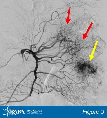
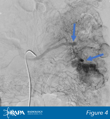
An angiogram shows mass (yellow arrow) in the left upper quadrant. Fine, tortuous vessels suggest tumor vascularity (red arrow). An early draining vein is also present (blue arrow).
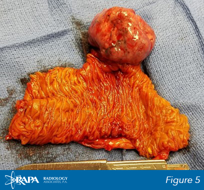
Given the location of the mass, the patient was sent to surgery and the mass was removed.
The exophytic mass is seen on the antimesenteric border of the surgical specimen. The mass has eroded into the bowel lumen (black arrow). (Figure 5)